Cyanobacterial cell division
Segmentation and tracking of individual cells and filaments to study growth, division, and differentiation.
Read more →Cesar Valades-Cruz
Advanced microscopy and image analysis for quantitative cell biology.
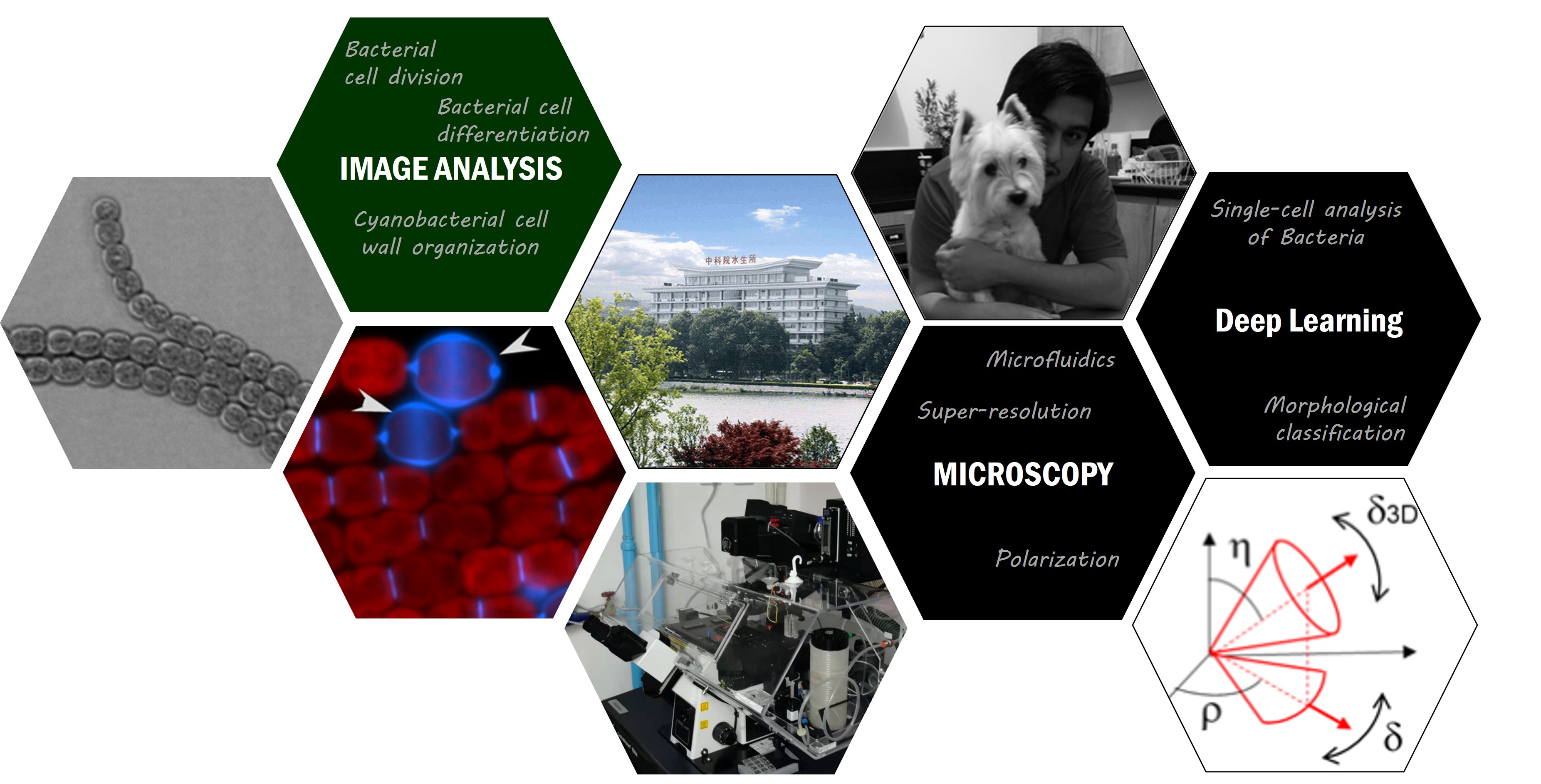
I develop microscopy and image analysis methods to study cell organization across scales. Explore the three areas below for an overview of the questions, methods, and projects.
Segmentation and tracking of individual cells and filaments to study growth, division, and differentiation.
Read more →Measuring molecular orientation and nanoscale structure using polarized super-resolution microscopy.
Read more →Navigation, tracking, restoration, and data workflows for complex microscopy datasets.
Read more →My current work at the Institute of Hydrobiology centers on cyanobacterial cell division. Collaborations in polarization imaging and computational microscopy extend these methods to molecular organization and live-cell data.
Funding & collaborations
Selected projects and programs in which I participate. See the CV for the full record.
Applications of Biophotonics and Image Processing in Microbiology · W2632090
High-throughput screening technologies for endosymbiosis.
Three-dimensional regulation of cell division in multicellular cyanobacteria · 32330003
Establishing cell function editing technology based on endosymbiosis · 32350028
Polarization microscopy for imaging of membrane organization.
Image-guided navigation and visualization of large live-cell microscopy datasets.
Data assimilation and lattice light-sheet imaging for endocytosis and exocytosis modeling.